All Departments
- Cataract Surgery
- Comprehensive Eye Tests
- Ear Conditions
- Eye Surgeries
- Facial Paralysis
- Head & Neck Surgery
- Laryngology
- Micro Ear Surgery
- Nose Conditions
- Nose Surgery
- Paediatric ENT
- Sleep Apnea & Snoring
- Throat Conditions
- Thyroid Diseases & Surgery
- Vertigo
- Vision Tests
Emergency Cases
888 666 4462Opening Hours
- Monday - Saturday: 9.00 am - 2.00 pm, 5.00 pm - 9.00 pm

Comprehensive Eye Tests
Comprehensive Eye Tests
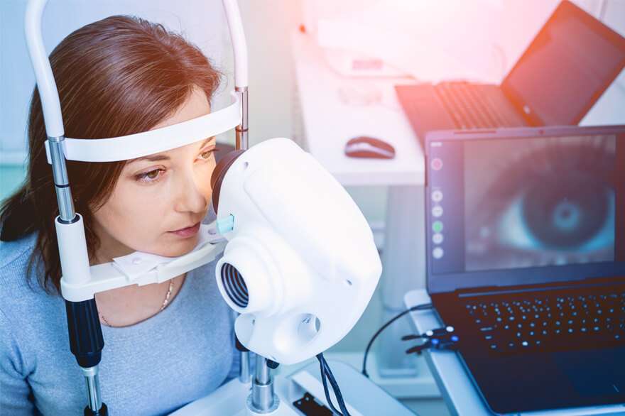
Regular, comprehensive eye testing is very important for maintaining healthy eyes and your best possible vision. Even if you feel that your vision is fine, there are eye disorders and diseases that can lead to vision loss if they are not detected early. A comprehensive eye testing can reveal significant details about your overall health condition.
What a Comprehensive Eye Exam Includes
You might be quite familiar with vision screening, which consists of reading letters from an eye chart to get a quick assessment of how clearly you can see. A comprehensive eye exam is quite different from a vision screening test, as it includes a range of tests that evaluate your vision and overall eye health thoroughly to help identify eye diseases and disorders.
The full range of tests the eye doctor recommends depends on various factors, but the following are the common tests done at our hospital:
Patient History
Analyzing your health history is an important part of a comprehensive eye exam. It’s important that your eye doctor knows your health history because it relates to eye or vision symptoms, health conditions you or your family members have experienced, your allergies, the medications you take, and your work or environmental conditions.
Factors of your health history can indicate you’re predisposed to certain eye diseases or disorders. Additionally, they can indicate any specific interventions that may be necessary to help keep your eyes healthy and your vision clear.
Visual Acuity
The visual acuity test, in which you read letters from a Snellen Eye Chart, is the most notable part of a comprehensive eye exam.
As you move down the chart the letters become smaller. Once your eye doctor determines the smallest line of letters, you’re able to read, the doctor will present the results with the familiar ratio of 20/20, 20/40, etc. The ratio will indicate the distance at which you can see an object from 20 feet away compared to the distance at which a person with normal visual acuity could see that object. For example, a 20/60 ratio indicates you can see an object from 20 feet away that someone with normal vision could see from 60 feet away.
While the results from the visual acuity test are an initial indicator that corrective lenses may be necessary, it doesn’t provide all the necessary information to determine what your treatment might be.
Refraction
A refraction test determines your corrective lens prescription. During the test, you are most likely to look through a phoropter – looking instrument with multiple lenses and dials commonly found in an eye doctor. Using the phoropter, your eye doctor will present a series of lenses of differing power and ask you from which one do you see better. Based on your answers, your doctor will continue to narrow down the options until reaching a final lens that best compensates for your refractive error (such as nearsightedness, farsightedness, astigmatism, or presbyopia).
Eye Movements
Visual acuity testing and refraction analyze your vision when looking at an immobile target. But the eyes have to move, focus, and work together to see a single, clear image. This particularly applies when shifting your focus from one object to another or when following a moving object.
During a common eye test used to analyze eye movement, the doctor will ask you to hold your head still and use your eyes to slowly follow a hand-held target. Disorders with eye movements can affect reading, participation in sports, and other activities. Severe eye movement disorders may indicate a condition such as amblyopia (lazy eye) or strabismus (cross-eyed).
Eye Pressure Test
Glaucoma is among the leading causes of blindness. Vision loss happens when the intraocular pressure (IOP) inside the eye is very high, causing damage to the optic nerve. Monitoring IOP on a regular basis is crucial for the early detection of the disease.
Tonometry is a test that measures the pressure inside your eye referred to as IOP. During this test, the eye is numbed with eye drops and a device called a tonometer gently touches your eye to measure the intraocular pressure. The procedure lasts just a few seconds and is painless. The instant results allow the doctor to establish if further glaucoma testing is required.
There are several other methods too for testing IOP, such as non-contact tonometry, electronic tonometry and Schiotz tonometry.
Pupil Dilation
Under normal circumstances, it’s difficult to see the back of your eye, as this area houses important structures such as your optic nerve and macula and a system of blood vessels that supply your eye. To obtain the best view, doctors have to dilate your pupils so they will remain enlarged to facilitate a thorough examination.
Your eye doctor will put dilating eye drops in both your eyes. After about 20 or 30 minutes, you’ll find it difficult to focus on objects up close, and you’ll become more sensitive to light, especially sunlight. Before you leave the clinic, you need to wear the disposable sunglasses available at the hospital or bring your own sunglasses. The effects of the drops will diminish in a few hours.
Slit Lamp
A slip lamp is one of the important and most useful devices the eye specialists use for examining your eyes. The slit lamp consists of a microscope with a high-intensity light source (i.e., a slit) that enables the doctor to more precisely assess the external and internal structures of your eyes for damage or pathology.
Eye doctors can thoroughly examine the front part of your eye to diagnose conditions as:
Cataracts happen to be one of the most widely existing eye problems. Light passes through a clear eye lens to your retina (just like a camera), where images are processed. With cataracts affecting your eye lens, light cannot pass through to the retina smoothly enough.
Signs and Symptoms of Cataracts
- Blurred, clouded or dim vision
- Problem seeing at night
- Problem seeing through light and glare
- Seeing ‘halos’ around lights
- Frequently changing contact lens prescription or eyeglasses
- Faded view of color
Dry eye is a condition of eye in which a person’s eyes does not produce enough tears or when the tears evaporate too quickly.
Symptoms
- A burning, scratchy or stinging sensation in eyes
- Eye redness
- Sensitivity to light
- Mucus production in or around the eyes
- Blurred vision
- Eye fatigue
- Issues in wearing contact lenses
Keratoconus is a vision disorder in which your eye’s cornea is unable to hold its round shape and becomes thin and irregular (cone) shaped.
Symptoms
- Double vision while looking with just one eye
- Objects both near and far looks blurry
- Light streaks
- Triple ghost images
- Blurry vision that makes it hard to drive
A corneal abrasion is a scratch on the front of your eye (cornea). It can happen if you poke your eye or something gets trapped under your eyelid. Most people can recover completely within 3 days. Symptoms may include eye pain, redness, sensitivity to light and a feeling like a foreign body is in the eye.
Conjunctivitis, commonly known as “pink eye,” is an infection that happens when the conjunctiva is irritated by an infection or allergies. It may result in red, itchy and painful eye.
When the pupils are dilated, the doctor can also assess your retina health (the back of the eye), including your macula and optic nerve. This part of the eye examination facilitates the diagnosis and assessment of conditions that can cause serious vision impairment or blindness, if left untreated, such as::
Diabetic Retinopathy is an eye disorder that can develop if you are diabetic and your blood sugar is not under control. High blood sugar levels can damage blood vessels of the retina in your eyes. In this condition, the blood vessels swell, leak fluid and blood into the retina.
The macula is the central area of the retina that helps you see greater detail and provides the best color vision. The macula tends to break down as you age. This is called age-related macular degeneration (ARMD).
There are 3 types of procedures for repairing a damaged or detached retina, which sometimes may be used in combination.
• Pneumatic retinopexy
• Scleral buckle
• Vitrectomy
Additionally, the blood vessels status in the back of your eye can provide early indications of overall health problems, such as diabetes or high blood pressure, often before you are aware of them. This is yet another reason to get a comprehensive eye test done on a regular basis.
Meet Our Doctors

Dr. Aravind Kumar Alwala
MBBS (Osm)., MSENT specialist, Chief Endoscopic Sinus Surgeon
Make an Appointment
Dr. Laxmi Priya Pallapolu
MBBS (Gold Medalist)., MS Ophthal (FICO)Consultant Ophthalmologist, Chief Phaco Surgeon
Make an Appointment